REMS vs DEXA: Doctors Explain Bone Density Testing
Summary
Many people assume a newer bone density test must be better than the old one. This video-driven deep dive explains why that is not automatically true for REMS (radiofrequency echographic multispectrometry). REMS uses ultrasound signals plus algorithm-based pattern analysis to estimate bone density, producing T-scores and Z-scores like a DEXA scan. The big advantages are no radiation, portability, and potential for more frequent monitoring. The key limitation is that REMS is calibrated against DEXA, so it cannot be more accurate than the standard it is trained on. Practical takeaways help you discuss options with your clinician.
🎯 Key Takeaways
- ✓REMS is an ultrasound-based approach that analyzes raw signal data, not just ultrasound images, to estimate bone density.
- ✓Its headline benefit is zero ionizing radiation, plus portability and potential point-of-care access.
- ✓DEXA radiation exposure is typically very low, the video compares it to roughly 5 to 10 transatlantic flights, so radiation is not usually the main barrier.
- ✓Because REMS is calibrated against DEXA datasets, it cannot be more accurate than DEXA, at best it can be close or equivalent.
- ✓REMS may be especially appealing where DEXA access is limited or when more frequent monitoring is desired, but coverage and availability vary.
What most people get wrong about “new” bone tests
The most common mistake is assuming that a newer bone density test must be superior simply because it is newer.
This video’s perspective is more skeptical and more useful: bone testing is a journey of tradeoffs. A test can be exciting, portable, and radiation-free, and still not outperform the long-standing standard.
That framing matters because bone density testing is not just about getting a number. It is about getting a number you can trust, compare over time, and use in real-world clinical decisions. For decades, DEXA (also called DXA) has been the “gold standard” largely because it has been studied extensively, validated, and woven into guidelines and fracture-risk tools.
At the same time, the discussion highlights why people keep asking about alternatives: access, convenience, cost, and the desire to avoid even tiny amounts of radiation.
Did you know? A DEXA scan uses a very small dose of ionizing radiation, and major radiology organizations describe it as low exposure compared with many other imaging tests. See the patient-friendly overview from the Radiological Society of North AmericaTrusted Source.
What REMS is (and why the name confuses everyone)
REMS stands for radiofrequency echographic multispectrometry.
If that name makes you think of radio waves, you are not alone. The video spends time clearing up a key confusion: “radiofrequency” in this context is not meant the way most people use it in everyday life (electromagnetic radiation like radio, Wi-Fi, or light). Instead, REMS is built on ultrasound, which is a mechanical wave that travels through a medium such as tissue and bone.
What is unusual here is not that ultrasound is being used near bone. It is that the device focuses on the raw signal information coming back from the ultrasound transducer, then uses complex processing (often described as AI or algorithmic pattern recognition) to connect that signal to bone status.
Historically, the conversation positions REMS as a technology that originated in Italy in the early 2000s and received European regulatory clearance (CE marking) in the mid-2000s. Uptake in the United States and Canada is described as slower.
Important: Availability and coverage vary widely. Even if a test exists, it may not be covered by your plan or available in your region, so it is worth asking your clinic what they offer and why.
REMS vs “regular ultrasound”
Standard medical ultrasound typically turns the returning signal into an image you can interpret visually. REMS, as described here, is more like, “Give us the raw sound information, and we will interpret patterns in it.”
That distinction is the heart of the video’s “journey of discovery” angle. It is not just a different machine, it is a different philosophy of measurement.
How REMS works, explained like a physics demo
The explanation in the video is refreshingly concrete.
An ultrasound system uses a transducer that behaves like a speaker and microphone combined. It sends out ultrasound waves, those waves interact with tissues and bone, and echoes return to the transducer. The device converts those echoes into an electrical signal (through a piezoelectric effect), then software processes that signal.
In conventional ultrasound, processing is aimed at producing an image. In REMS, the emphasis is on the signal itself. The approach described is essentially: skip the image-building step, analyze the raw returning “radiofrequency” signal from the ultrasound system, and let an algorithm map signal patterns to bone characteristics.
A key detail is that REMS does not rely on a single frequency. It uses a pulse containing multiple frequencies, then performs spectral analysis (breaking the signal into its component frequencies and examining the “power” at each frequency). The underlying idea is that bone’s structure affects how sound is absorbed, transmitted, and reflected. The algorithm then compares those patterns to reference patterns.
This is where the video’s metaphor lands: it is almost like “looking down a femur and yelling ‘Hey,’ then listening to the echo,” and using that echo to infer bone quality.
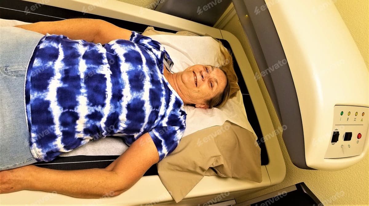
What the test looks like for a patient
The practical walkthrough is simple and reassuring. The scan is typically done at the hip (often both hips) and the lower back, similar target sites to DEXA.
Just like DEXA, the output includes a T-score and a Z-score.
If you have questions about how these scores are used in diagnosis and risk assessment, reputable patient summaries are available through the Bone Health and Osteoporosis FoundationTrusted Source.
Why consider REMS if DEXA already exists?
This is the part of the discussion that feels most practical: if DEXA is well-studied, why care about REMS at all?
The first advantage is straightforward. Because REMS is ultrasound-based, it involves no ionizing radiation.
The video adds an important nuance, though. DEXA radiation is already quite low. Patient-facing radiology resources describe DEXA as a low-dose exam, and the video uses a memorable comparison, roughly equivalent to several long flights. In other words, “no radiation” is a real advantage, but it is not necessarily the only deciding factor for most people.
The second advantage is access and portability. The discussion emphasizes that REMS can be a point-of-care style device, smaller and more portable than a large DEXA machine. DEXA also requires a facility setup that accounts for radiation protection, which adds cost and complexity.
The third advantage is potential cost per test. The clinicians discuss that REMS may be cheaper to perform, although real-world out-of-pocket cost can be unpredictable if insurance does not cover it.
The most intriguing advantage in this video is frequency. Because there is no radiation, the concept of testing more often becomes more plausible. The discussion gives examples like doing checks every three or six months in situations where someone wants closer monitoring.
Pro Tip: If your goal is to track change over time, ask the clinic how they ensure repeatability, meaning the same site, same positioning, and comparable technique each time. Consistency can matter as much as the device.
When “more frequent testing” might come up
This is not a suggestion to test constantly. It is a reminder that different clinical situations create different needs.
For context on how osteoporosis is typically assessed and monitored, including the role of bone density testing, see the overview from the National Institute of Arthritis and Musculoskeletal and Skin DiseasesTrusted Source.
Is REMS “better” than DEXA? The calibration reality check
The video asks the question many viewers are thinking: could REMS be more accurate than DEXA?
The answer given is a clear “no,” at least not right now, and the reasoning is worth understanding.
This perspective emphasizes calibration. REMS is trained and validated by comparing its readings to DEXA results. If DEXA is the reference standard used to label “normal” vs “abnormal” bone density, then REMS cannot logically exceed the accuracy of the standard it is anchored to. At best, it can approach DEXA’s performance, and in practice it will have its own sources of error.
That does not make REMS useless. It simply reframes it as a tool with different strengths. A test can be “good enough” for certain use cases, especially where DEXA is unavailable, and still not replace DEXA in guideline-based care.
The video also points out a real-world issue that affects many ultrasound-based measurements: operator dependence. How the probe is placed, the pressure applied, and the exact site measured can introduce variability. The optimistic note is that improved algorithms and better hardware may reduce that variability over time.
What the research shows: DEXA remains the widely accepted standard for diagnosing osteoporosis and monitoring bone density in many guidelines, largely because it has extensive validation and standardized reference databases. Patient summaries from RadiologyInfo.orgTrusted Source reflect this clinical role.
Expert Q&A
Q: If REMS has no radiation, should I switch from DEXA automatically?
A: Not automatically. A radiation-free test can be appealing, but what matters is whether the result is reliable, comparable over time, and accepted by your clinician for decision-making.
If you already have DEXA results, staying with the same method can make trending clearer. A reasonable next step is to ask your clinician whether REMS results at your facility are validated, how repeatable they are, and how they would use the result in your care plan.
Dr. Paul Zalzo, MD
Expert Q&A
Q: Could REMS ever become the new gold standard?
A: It is possible for technologies to evolve, especially as algorithms improve and operator variability is reduced. But becoming a gold standard usually requires large, long-term studies, standardized protocols, and broad adoption so results are comparable across clinics.
For now, it is best viewed as a promising option to watch, particularly for improving access where DEXA is hard to obtain.
Dr. Brad Weining, MD
A practical, step-by-step way to decide what to ask for
You do not need to become a physicist to make a good decision. You just need a plan for the conversation.
How to talk to your clinician or imaging center
Start with your goal, not the gadget. Are you screening because of age or risk factors, confirming a diagnosis, or monitoring change over time? The best test is the one that answers your specific question.
Ask what tests are available locally. Many regions still primarily offer DEXA. If REMS is offered, ask where it is performed (clinic vs hospital) and who performs it.
Ask how results will be used. Will the number feed into a fracture-risk calculation? Will it change treatment decisions? If the answer is “probably not,” it may not be worth paying out of pocket.
Ask about repeat testing and trending. If you are planning follow-up measurements, ask whether the same device and protocol will be used each time, and what time interval is typical for your situation.
Ask about cost and coverage upfront. The video notes that insurance coverage may lag behind newer tools. A quick call to your insurer can prevent surprise bills.
»MORE: Consider keeping a one-page “Bone Health Snapshot” for appointments, including prior DEXA dates, T-scores by site (hip, spine), fracture history, and current medications or supplements. It makes follow-up discussions faster and more accurate.
A quick checklist to bring to the appointment
A broader, evidence-based overview of osteoporosis prevention and management is available from the U.S. National Library of Medicine (MedlinePlus)Trusted Source.
Key Takeaways
Frequently Asked Questions
- What does REMS stand for in bone density testing?
- REMS stands for radiofrequency echographic multispectrometry. Despite the name, it is an ultrasound-based method that analyzes returning signal patterns to estimate bone density.
- Does REMS use radiation like a DEXA scan?
- No. REMS is ultrasound-based and does not use ionizing radiation. DEXA does use a small amount of ionizing radiation, but it is generally considered low dose by radiology organizations.
- Can REMS be more accurate than DEXA?
- In the video’s framing, not currently. REMS is calibrated against DEXA reference data, so it cannot exceed the accuracy of the standard it is trained to match, though it may approach it in some settings.
- What body areas are typically scanned with REMS?
- The video describes scanning the hip (often both hips) and the lower back, similar to common DEXA sites. The scan uses gel and a probe and usually takes about 10 to 15 minutes.
- Will REMS results include a T-score and Z-score?
- Yes, REMS can produce T-scores and Z-scores like DEXA. A T-score compares to a young adult reference, while a Z-score compares to an age-matched reference.
Get Evidence-Based Health Tips
Join readers getting weekly insights on health, nutrition, and wellness. No spam, ever.
No spam. Unsubscribe anytime.